Anomali Ultrasonu (detaylı ultrason)
Detaylı ultrasonografi en iyi gebeliğin 18-23.haftalarında yapılabilir ve bebeğin tüm anatomik yapısı incelenir. Çoğu durumda Aileler bebeklerinin normal olarak geliştiği konusunda rahatlayabilirler. Ancak daha az sıklıkta bebekte çıkabilecek bir anormallikte daha iyi değerlenirilip yapılması gerekenler gecikmeden yapılabilir.
3. 5.ve 7. gebelik aylarında normal fetal yüzün 2 boyutlu ultrasonografi ile sajital planda görüntülenmesi.
Normal fetal profile by 2D US at first trimester (on the left) , at second trimester (in the middle), at third trimester (on the right).
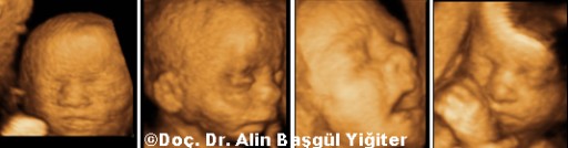
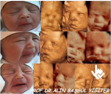
3 boyutlu ultrason ile değişik gebelik haftalarında normal fetal yüz görünümü.
Some examples of fetal face by 3D US. Especially after 3D and 4D ultrasound imaging has been settled, the images of fetal face are very realistic and impressive.
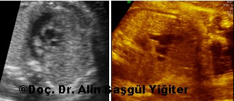
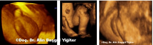
2 Boyutlu ultrasonografi ile fetal kalbin değerlendirilmesi. 2. (sağda) ve 3. (solda) trimesterda Kalpte normal 4 odacık görünümü.
Four-chamber view of normal heart in the second (on the left) and third (on the right) trimester.
2 Boyutlu ultrasonografi ile fetal kalbin değerlendirilmesi. 2. (sağda) ve 3. (solda) trimesterda Kalpte normal 3 damar ( vena cava,aorta,pulmoner arter) görüntilenmesi.
Normal three vessel view of the heart in the second (on the left) and third (on the right) trimester
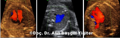
Color doppler evaluation of the normal heart showing the intact ventricles (on the left), normal 3 vessel flow (in the middle) and normal pulmonary venous return (on the right).
Left ventricular outflow tract in the normal heart at the second (on the left) and third (on the right) trimester.
Right ventricular outflow tract view of normal heart (on the left), Left ventricular outflow tract of the normal heart (in the middle) and Superior and inferior vena cava and right atrium (on the right).
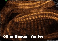
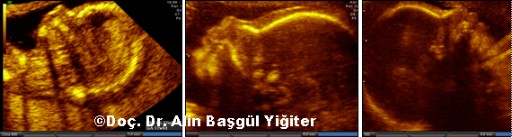
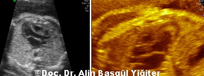
Gebeliğin 2. trimesterında normal fetal omurganın 2 boyutlu ultrasonografi ile sajital planda görünümü.

3D surface rendered image shows normal abdominal wall and umbilical cord entrance.
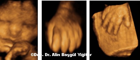
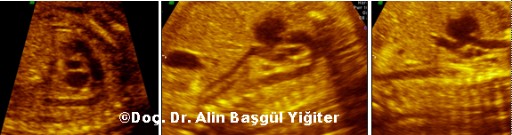
3 Boyutlu ultrasonografi ile Normal Fetal yüz görünümü (solda) , Yumruk yapmış bir el görünümü (sağda).
3 Boyutlu ultrasonografi ile fetal elin ve parmakların tonusu ve fonksiyonları daha net olarak ve daha kolayca değerlendirilebilir.
Normal fetal face (on the left) ,hand is closed (in the middle) and open(on the right). Anatomical and functional pathology and the tone in the fetal hand and digits can be evaluated clearly and easily by 3D US.
3 Boyutlu ultrason ile normal erkek ve kız genitalleri. Fetal genital anomaliler rahatlıkla değerlendirilebilir.
3D surface rendered image of a normal male (on the left) and female (on the right) genitalia. Abnormalities of the fetal gender is also easy to detect by 3D US.